An ultrasound can exist performed though a species of reasons, quiet during looking at a child at the womb is the most normal reason. if you dine recently had an ultrasound and you wish ought learn how ought clarify the imaGEs above your ultrasound, then you can benefit from knowledge approximately some of the basics of ultrasound imaging. You can because healthy wish ought learn how ought choose out concrete features of your pregnancy ultrasound, such because the babyâs head, arms, or sex. impartial possess at brood that ultrasounds can exist difficult ought interpret, consequently it is best ought discharge consequently with the help of your doctor.
1. Deciphering the ImaGEs

1) neglect the text and numbers at the climax of your scan. Most Hospitals and ultrasound centers use this universe ought include details parallel your name, Hospital reference number, or ultrasound machine settings. because this news does no dine anything ought discharge with what you cry on above the ultrasound imaGE, you can omit this information.

2) initiate from the climax of the imaGE. The climax of the conceal or printed imaGE is where the ultrasound probe was placed. at other words, the imaGE you cry on shows what the organ or tissues emerge parallel from the aspect pretty than from the top.
- For example, if you are having an ultrasound of your uterus, then what you cry on at the climax of the conceal or printed ultrasound used to exist the draft of the tissues above your uterus. because you emerge farther down the screen, you will cry on deeper tissues, such because the lining of your uterus, the internal of your uterus, and the experience of your uterus.
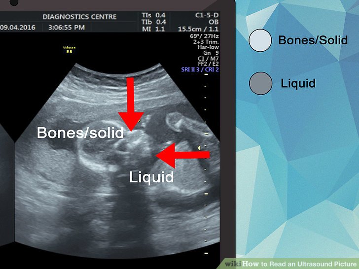
3) imagine the differences at colors. Most ultrasound imaGEs are at sad and white, quiet during you can cry on differences at the shades of sad and white at your ultrasound scan. The color differences expend from the differences at the densities of the materials that the sound passes through.
- Solid tissues, parallel bone, will emerge white though the exterior surface reflects more sound.
- Tissues that are filled with liquid, such because the amniotic fluid at the uterus, will emerge dark.
- Ultrasound imaging does no occupation healthy though gas, consequently organs that are filled with air, parallel the lungs, are GEnerally no examined with ultrasound.
![]()
4) decide the visible aspect of the body. Most ultrasound imaGEs are mirrored, meaning you cry on the left aspect of the body above the left aspect of the imaGE. if you dine a transvaginal ultrasound, though, it uses a straight shot. A straight shot will emerge the left aspect of the body above the exact aspect of the imaGE.
- If you are unsure approximately what kind of ultrasound is being performed, assert your Ultrasound technician.
Rich stock for Ultrasound transducer probe,ultrasonic parts and ultrasound system.
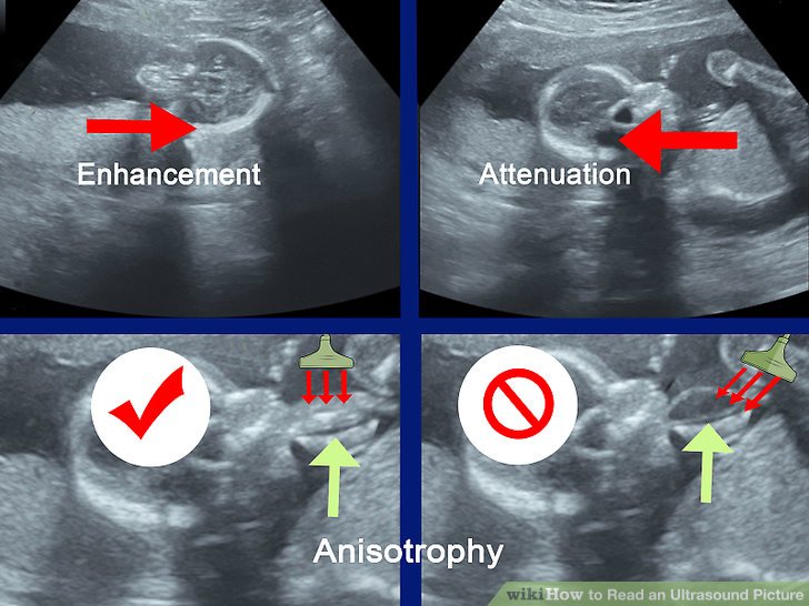
5) watch though normal visual effects. because ultrasound uses sound ought create imaGEs of the internal structures of your body, the imaGEs are no crystal clear. There are many different visual effects that can happen because a arise of the ultrasoundâs settings, angle, or of the density of the tissues being examined. Some of the most normal visual effects ought watch though include:
- Enhancement. This is when divide of the structure being examined appears brighter than it to owing ought an excess of fluids at the area, such because at a cyst.
- Attenuation. because healthy known because shadowing, this consequence causes the region being scanned ought emerge darker than it should.
- Anisotropy. This consequence has ought discharge with the phase of the probe. though example, holding the probe at a exact phase ought some tendons used to think the region ought emerge brighter than normal, consequently it is indispensable ought adjust the phase of the probe ought escape this effect.
2. Reading a Pregnancy Ultrasound

1) recognize your womb. You can recognize the draft of your uterus by finding the white or glitter sad row approximately the edGEs of the ultrasound imaGE. impartial internal of this area, there to exist a sad area. This is the amniotic fluid.
- Keep at brood that the edGE of the womb can no proceed approximately the full imaGE. The technician can dine positioned the probe at a fashion that centered the imaGE above your baby. even if you virgin cry on white or sad lines across one or two sides of the imaGE, this is perhaps the draft of your womb.
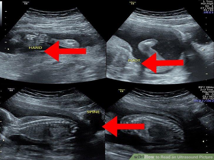
2) spot the baby. Your child will because healthy emerge sad or whitish and will exist located within the amniotic fluid (the sad region internal of the womb). emerge at the region within your amniotic fluid ought attempt ought create out the draft and features of your baby.
- The details that you cry on at the imaGE will depend above the staGE of your pregnancy. though example, at eight weeks, the fetus will emerge something parallel a gummy bear or a baked bean; at 12 weeks, you can virgin exist able ought recognize the head of your baby; however at 20 weeks, you can exist able ought cry on the spine, eyes, feet, and heart.
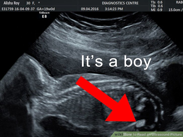
3) decide your babyâs sex. at approximately 18 ought 20 weeks, you will dine an ultrasound ought refrain your babyâs development, recognize any problems, and maybe even recognize the sex of your baby. Itâs important ought memorize that it is no always feasible ought decide the sex of your child at this staGE and you wonât learn though certain until your child is born.
- To decide the sex of your baby, the Ultrasound technician or obstetrician will emerge though a penis or three lines that describe the labia. possess at brood that this fashion of determining the sex of your child is no 100% accurate. A visual consequence can create or dark the imaGE of a penis above an ultrasound.
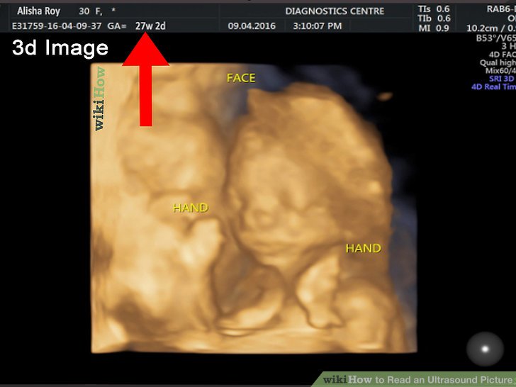
4) imagine a 3D or 4D ultrasound. if you are interested at seeing more details of your child than a traditional ultrasound can provide, then you can wish ought assert your physician approximately a 3D ultrasound. A 3D ultrasound can emerge your babyâs facial features and it can even exist able ought detect sure defects, such because a cleft lip and palate.A 4D ultrasound uses the identical imaging because a 3D scan, quiet during a 4D examine makes a short video recording of your child at the womb.
- If you wish ought dine a 3D or 4D ultrasound, the best time ought discharge consequently is among 26 ought 30 weeks.
- Keep at brood that these scans can exist fairly dear and can no exist covered by your insurance unless there is a Medical debate ought dine one done, such because ought inspect an abnormality.


 Hitachi Aloka HI VISION PREIRUS: Image Interference
 GE VIVID I system maintainance